Contents |
Corneal Neovascularization
Introduction
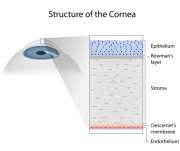
The cornea is the clear, dome-shaped structure that covers the iris (where the color is) and is the first surface light strikes on its way into the eye to the retina. Normally, the cornea is very clear, with an even, smooth surface. It is clear because the layers of tissue are in alignment and because it is a relative dehydrated tissue; the skin cells lining the back surface, called the endothelium, are constantly removing extra water from the cornea. (The water pumped out of the cornea mixes in with the aqueous humour, the liquid that fills the front part of the eye, then the excess liquid drains away.)
All tissues of the body require oxygen for their proper metabolism. The oxygen enters the body through the lungs, of course, and then passes through the lining of the lungs and enters the circulatory system. From there, it is carried to the rest of the body.
But the cornea is an avascular tissue, meaning it has no blood vessels. Blood vessels growing in the corneal tissue would compromise its transparency, so it must get its oxygen in other ways, through absorption from the tear film and the atmosphere. (Oxygen also comes up the edges of the cornea, in the tiny arteries of the conjunctiva; but once those vessels reach the area where the sclera becomes the clear cornea, they reverse direction and become veins.)When the cornea is deprived of enough oxygen the endothelium pump loses efficiency and is unable to maintain the relatively dehydrated state of the corneal tissue, and therefore it begins to swell. When the decrease in available oxygen becomes chronic, the cornea responds by allowing tiny new blood vessels to grow. This is corneal neovascularization.
In the early days of contact lenses, materials did not allow oxygen to pass through at all, and CN was quite common; some eyecare practitioners accepted CN as being a “normal” result of contact lens wear, because almost every patient had it after a short time of using their lenses. Now, blood vessels growing in the tissue of the cornea should never be considered normal, but are always a sign that the level of oxygen getting to the cornea is not adequate.
These new blood vessels are abnormal, not only by where they are growing, but they also are unusually sized and shaped, and are very fragile. They begin to grow from the edges of the cornea and progress towards the center, where they begin to interfere with vision.
Causes of Neovascularization
There are several known causes of corneal neovascularization (CN); the most common one is contact lens overwear. Overwear can be the result of wearing contacts too many hours in a day, sleeping without removing non-extended wear lenses, using contacts that are no longer transmitting oxygen like they did when new, and allowing the contact lenses to become dirty or to form deposits on their surfaces from inadequate lens care.
Inflammation is another source of CN as the result of trauma or injury, and from blepharitis, uveitis, or keratitis. (In medical terms, the suffix “-itis” added to the name of the structure means that structure is inflamed. Inflammation is characterized by swelling, redness and pain, and is not the same as an infection, which is caused by micro-organisms.) Corneal ulcers can cause CN, as can glaucoma and other ocular surface diseases like rosacea or lupus.
People who are at risk for CN are
- Contact lens wearers, especially those who are non-compliant with lens care
- People who have large amounts of nearsightedness (myopia)
- Sufferers of dry eye disease
- Those who have an ocular surface disease (rosacea, lupus)
- Those who have an underlying inflammatory disease (multiple sclerosis, Crohn’s Disease)
- Those with untreated, or inadequately treated, infection of any ocular tissues
Symptoms and Signs
People who are developing or who have already developed corneal neovascularization are not always aware of it. Symptoms can be subtle, like contact lenses not being as comfortable as they once were, or hazy vision.
It is usually the eyecare practitioner, using an instrument known as a slit lamp to look at the contact lenses and the corneal surface under them, who first recognizes that the cornea is no longer completely clear, and that there are vessels invading where they should not be. The new vessels are very tiny and cannot usually be seen by the naked eye, but the slit lamp has a bright source of light and magnification so the tissues can be seen clearly and easily. A slit lamp examination of the surface of the eyes is part of a routine vision exam, as well as for evaluating contact lenses.
Treatment
Corneal neovascularization is treated by doing whatever is necessary to increase the amount of oxygen that is reaching it.
Any existing inflammation of the ocular tissues must be treated; however, most CN has a more obvious cause, that of contact lens overwear.
Contact lens overwear is not seen as much now as it once ways, because of new technology in materials, lens designs and the ability of newer lenses to transmit oxygen at a much higher level than ever before. However, even though it is more unusual in the present, it does still exist, usually in patients still wearing the same type of contact lenses they were originally fit with when they first began to use them.
Contact lens wearers may need to be refitted into a new lens made from a material like silicone hydrogel which transmits oxygen at almost the same level as having no lens present on the eye at all. Usually, this alone will solve the problem, but some patients may need to reduce their daily wearing time, switch to an extended-wear type of lens but wear it only during the day, and, in very extreme cases, to discontinue contact lens wear completely.
It is important to understand that once the corneal endothelial pump begins to break down, it will almost never return to its pre-CN state. The new blood vessels will never go away again; they will empty and no longer carry blood into the cornea, but they are still there. (These are called “ghost vessels,” because without blood in them, they look a little ghostly, being clear and hard to see.)
If left to grow, the new blood vessels continue further into the corneal structure, and as noted above, they are quite fragile. If one of them ruptures (a slight bump to the head, a coughing spell or a foreign body in the eye can all cause rupture of the vessels) the hemorrhage itself, being opaque, will cause visual symptoms and can even leave scar tissue in the cornea.
Light does not pass through tissue with blood leaking into it, nor does it pass through scar tissue except in a very limited way. The devastating loss of vision that can result should be obvious.
Prognosis
Once blood vessels have grown into the corneal tissue, they will not disappear completely. By increasing the oxygen getting to the cornea, it is possible to stop their growth, but the best result will still leave ghost vessels there. If at any time, the cornea is again compromised for lack of oxygen, those vessels will fill up and begin to grow again.
However, by getting regular contact lens checkups and following the proper wearing schedule, most patients can stop CN in its tracks.
If the cause is inflammation of ocular tissue from another direction, or an infection, the underlying cause must be treated.
Summing Up
Corneal neovascularization is never considered to be normal, and it should not be accepted as just one more result of contact lens wear.
As noted earlier, if left to grow, these vessels can cause major vision disruption.
Even if it is the result of inflammation unconnected with contact lens wear, it is possible to treat that condition with anti-inflammatory medications and other measures. While it is as yet not possible to cure diseases like rosacea, lupus or MS, we can get regular medical care and do what we can to minimize the influences they have on the entire body as well as the tissues and structures of the eyes. Sometimes, we just need to do the best we can.
Most corneal neovascularization is the result of contact lens wear that is compromising the amount of oxygen getting to the cornea. It is easy to stop CN when it is due to overwear or compromised oxygen transmissibility because of lenses that are not in optimum condition. All we have to do in this case is follow the instructions of the eyecare practitioner.


